Xray microscopy (XRM)
Overview
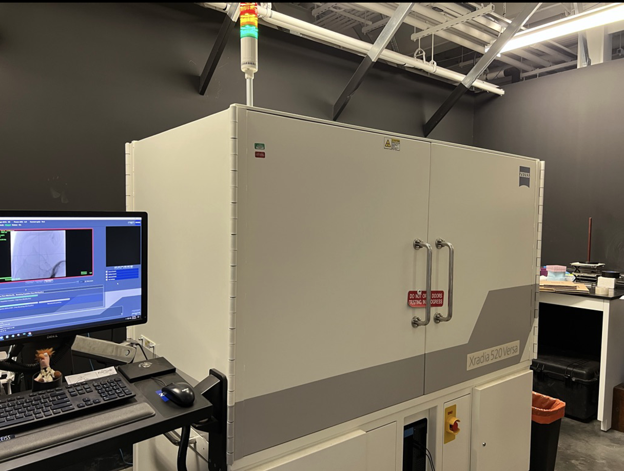
To extend our imaging capabilities, we added a ZEISS Xradia 520 Versa X-ray microscope (XRM) in April 2018, supported by our research collaboration with Valent BioSciences, Sumitomo Chemical Company, and the Donald Danforth Plant Science Center.
This is currently the only lab-based XRM in the world dedicated exclusively to studying plant science, and our February 2022 publication in Plant Physiology (Duncan et al., 2022) describes how we have used XRM for a wide range of economically important plant species.
How it works
The XRM is a powerful and versatile instrument for generating detailed 3D volume data of delicate and complicated samples. The multiscale capability allows high magnification region-of-interest scans to be situated within the context of lower magnification scans of entire samples, without removing the sample from the instrument. The animations and screen shots below illustrate both the range of plant and soil samples that we have studied as well as the multiscale capability of the instrument. We’re also using XRM to study arbuscular mycorrhizal fungi (AMF) and their interaction with roots in situ. We developed various strategies for generating contrast of AMF structures for in vitro and in situ visualization of hyphae, spores, arbuscules, and vesicles.

Root-microbe colonization
Roots of Brachypodium colonized by Rhizophagus irregularis, contrasted with osmium and imaged by XRM in situ.Soil aggregate
Multiscale overlaid scans of soil aggregate, part of a project to use X-ray imaging to evaluate pore space from soils under differing management strategies.Soil aggregate
Multiscale overlaid scans of soybean flower with ovary containing three ovules, surrounded by pollen-containing anthers. Polar nuclei and synergids are visible in the high magnification scan of a single ovule.